Diagnostics
MAGNETIC RESONANCE IMOGRAPHY
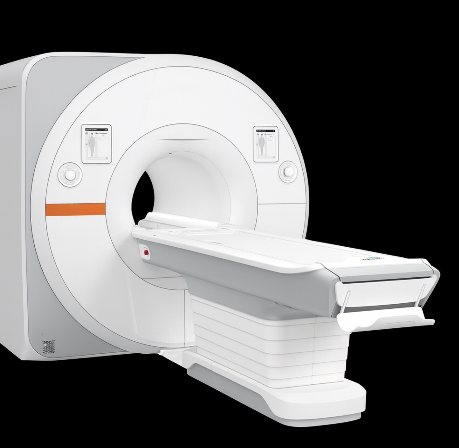
Magnetic resonance imaging (MRI), also known as nuclear magnetic resonance imaging, is a diagnostic procedure that has become widely used in recent years due to its excellent diagnostic capabilities and image quality. Unlike X-rays and computed tomography (CT), it does not use X-rays. The body part being examined is placed in a strong magnetic field, stimulating the tissue to emit measurable signals.
These signals are recorded and processed into an image using sophisticated calculations performed by powerful computers. Thus, unlike X-rays, an image of the body is not “taken” but rather “calculated.” This is the advantage of this examination method: the body segment can be electronically “sliced into sections.” This makes it possible to create a high-resolution, three-dimensional representation of the body’s interior, yielding far more information than with other examination methods. All soft tissue structures, such as muscles, tendons, ligaments, menisci, cartilage, fluids, and especially the intervertebral discs, are depicted with excellent clarity.
BONE DENSITY MEASUREMENT
Early detection and prevention are the best ways to protect against serious secondary diseases. One in three postmenopausal women is affected by bone structure disorders (so-called osteoporosis). According to the guidelines of the German Osteology Society (DVO), preventive bone density measurement is recommended for all women under 60 with one or more risk factors, as well as for all women over 60, all men and women with a history of bone fractures without adequate trauma, those with metabolic bone diseases (Paget’s disease, rheumatoid arthritis), and those undergoing long-term cortisone therapy (for example, for asthma). Dual-energy X-ray absorptiometry (DXA) is the safest preventive examination and the current “gold standard” according to the DVO guidelines. This method measures bone density at two different locations (lumbar spine and femoral neck). It uses low radiation and takes only about 10 minutes. This examination is also performed by Dr. Merettig and Dr. Stöber have arranged an appointment for you at the Diagnostic Center at Emser Platz.
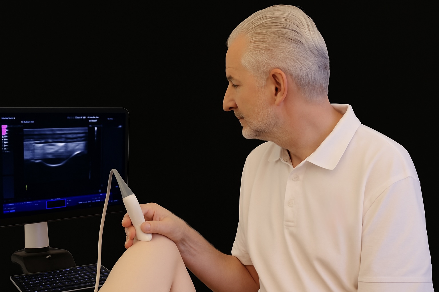
ULTRASOUND / SONOGRAPHY
Fast, straightforward, and completely safe: Ultrasound (sonography) allows for the detection of recent injuries, hematomas, joint effusions, bursitis, and tendon ruptures in the shoulder, hand, and knee, as well as rheumatic diseases. Its almost universal availability makes ultrasound extremely valuable for disease detection and follow-up examinations, particularly for muscle and tendon disorders and injuries.